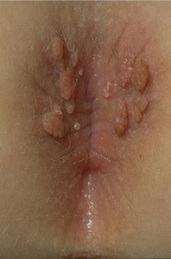
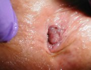
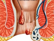
 Genital Warts are the clinical manifestation of an active infection of the male and female genital, perigenital and perianal region skin and mucous membrane, with certain types of human papillomavirus (HPV). The disease is both sexually and non-sexually transmitted (infected hands, iatrogenic, prenatal or perinatal transmission in children). The infection is very contagious and it is frequently associated to other sexually transmitted diseases (syphilis, HIV, etc.), which imposes the performance of the respective tests. In some countries, it is regarded as the most frequently encountered sexually transmitted disease. It should be mentioned that it could become malignant.
The histopathological examination of genital warts:
Only indicated in the cases that do not respond to or worsen during therapy (possible dysplasia), in the case of pigmented, infiltrated injuries or in the case of an uncertain diagnosis.
Condylomata acuminata are small cauliflower-like, whitish tumoral lesions, which tend to group in plaques. In women, they may be located at the level of the cervix or of the intravaginal mucous membrane (more rarely in men, at the level of the penile sheath). They can reach large forms, located in men at the level of the gland or perianally (more rarely in women).
Condylomata accuminata complications:
- microbial or fundal superinfections;
- malignant transformation (flat condylomata accuminata of the cervix, Cervical intraepithelial neoplasia (CIN), giant spinocellular penile condylomata).
Diagnosis explorations:
Mandatory
- tests to detect the simultaneous presence of other sexually transmitted diseases (syphilis, HIV infection).
Optional:
- Acetic acid 5% coating test, which, if positive, highlights a whitish network (used to detect subclinical forms, non-specific erythematous forms and to detect the extent of the infection around the obvious clinical injury area);
- Histopathological examination: detection of koilocytic-like cells. Only indicated in the cases that do not respond to or worsen during therapy (possible dysplasia), in the case of pigmented, infiltrated injuries or in the case of an uncertain diagnosis.
- Viral identification and viral staging using the PCR technique.
Treatment:
The choice of the optimum treatment method depends upon numerous factors: the location of the condylomata, for how long they have been present, the simultaneous infection of the partner, the immune status, the HPV subtype, other simultaneous genital infections, etc.
Local therapies: the local application of the Trichloroacetic acid, Podophyllin, Podophyllotoxin with condylomata destruction processes; cryotherapy, electro cautery, surgical or CO2 laser removal. Local immunomodulators and local antiviral mixtures or intralesional injections may be used.
General therapies: for extended, relapsing lesions, with an underlying severe immunodepression background, the treatment of the latent infection with HP, isoprinosine, retinoids, Interferon, immunostimulators, non-specific with Levamisole, BCG.
Hospital admission and guidance criteria:
The giant clinical forms, which do not respond to the applied therapies, occurring in the context of an underlying immunodepression.
Prevention measures:
Condoms: regardless of the treatment method used, the patient will be advised to use a condom for 3-6 months as of the clinical healing. Condoms do not offer full protection, as the infection can occur through the skin contact with the perigenital region. They do, however, ensure protection against the infection of the cervix with oncogenic HPV types.
The preventive and therapeutic vaccine therapy is currently under development (especially for HPV type 16, most frequently associated to cervix cancer).


